Ocular System In Horses – An Introduction
The eyes are an essential sensory organ. Fish, birds, insects and reptiles have them as well as just about every mammal. Finding food, defense, offense, mating or just having fun requires the brain to see what is around us.
It is interesting to see the embryonic development of eyes. They start as stalks of neural tissue sprouting from the front part of the brain. Reach straight out in front of you with both hands and pretend you are holding a ball. The arms are these stalks called the optic nerves and the cupped hands are the back of the eyeball called the retina. Your body represents the brain. In essence, the eyes are a direct extension of the brain visible to all who look at them.
Half of each eye’s nerves from the brain cross over to the other side to the opposite eyeball. The stalk develops forward into the eye socket of the skull and then forms a unique layer of light receptors called the retina. Unique to the horse is the shape of this and is called a ramped retina. This allows for a different focal point depending if the head is lowered for grazing (the flat of the face looking towards the horizon for predators) or raised to look down the nose at something close (you).
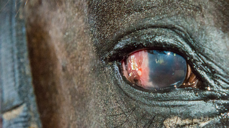
Following the retina forward is a lens to form uniform light rays. It is suspended in fluids and covered by a diaphragm (the uvea which contains the iris giving the color of the eye) to control the amount of light coming into the eye (the pupil). Unlike most animals, the horse adds a glob of tissue to this diaphragm to help shade the sun called the corpora nigra.
Finally the front of the eye is covered with a transparent membrane called the cornea. This membrane seals the inside of the eye from the outside and is fully transparent unless damaged. For further protection the eyeball is protected with upper and lower eyelids and a special 3rd eyelid called the nictitating membrane. The outside of the eyeball is bathed in fluid (tears) to lubricate, preserve corneal transparency, initiates the bending of the light rays for focusing and distribution of nutrition and immunoglobulins to maintain the health of the cornea. The tears are drained through small holes on the inside corner of the eye connected to a tunnel in the skull that empties the tears at the nostrils (the nasolacrimal ducts).
With so many parts to the eye, there are several areas where problems can occur. Trauma is the leading problem but infections, immune inflammation and neoplasia can also affect these structures. Loss of clear vision (corneal ulcer), loss of all vision (cataracts, blind), loss of the eyeball (enucleation) and tear overproduction or poor draining all can occur. Most of these can be seen with our own eyes especially if we are looking for them.
Ocular System In Horses – Blindness
Blindness In Horses
Ocular System In Horses – Cataracts
Cataracts In Horses
Ocular System In Horses – Corneal Ulcers
Corneal Ulcers In Horses
Ocular System In Horses – Enucleation
Enucleation is the removal of an eye ball. This is done when an eye is in the end stage of chronic inflammation or the active inflammation is too painful to leave the eye in.
Ocular System In Horses – Eye Injuries
Eye Injuries In Horses
Ocular System In Horses – Eye Medication Systems
Eye Medication Systems For Horses
Ocular System In Horses – Eyelid Wounds
Eyelid Wounds Of Horses
Ocular System In Horses – Glaucoma
Glaucoma In Horses
Ocular System In Horses – Nasolacrimal Ducts
The nasolacrimal duct (n-g duct) is a tube that runs through the skull starting at the inside corner of the eye and ends just inside the nostril.
Ocular System In Horses – Normal Eye Features
Normal Horse Eye Features
Ocular System In Horses – Discharges
Ocular Discharges In Horse Eyes
Ocular System In Horses – The Third Eyelid (Nictitating Membrane)
The Third Eyelid Of Horses (Nictitating Membrane)
Ocular System In Horses – Top 5 Eye Problems
This is a replay of a HorseTalk™ webcast. Be sure to enroll in the next one and join Doc T live.
Horse Immune Diseases – Eye Inflammation (Uveitis)
Eye Inflammation In Horses
Horse Neoplastic Diseases – Neoplasia In Eyes
Neoplasia In Horse Eyes
Responses